Single Cell and Spatial Omics
Gain spatial resolution of gene expression at tissue, cellular, and subcellular levels with advanced spatial genomics workflows.
At the core of our spatial transcriptomics capabilities are the 10x Genomics platforms: Xenium, Visium, and Chromium. Together, these platforms enable comprehensive, high-resolution insights into tissue structure and function for studies across virtually all research fields, including cancer research.
Integrate Molecular and Spatial Data for Advanced Discoveries
Spatial genomics is a revolutionary tool that lets researchers examine the expression of specific genes at the tissue and single-cell levels. This has significant advantages over previous methods, which homogenize diverse cell and tissue types, forcing researchers to draw conclusions based on averaged expression levels. Spatial transcriptomics platforms unlock the details of healthy and diseased tissues, offering insights into cell-cell interactions, the resident cell types of diverse tissues, and complex tissues like the tumor microenvironment and tertiary lymphoid structures.
Single-cell omics integrate spatial information to help researchers understand the activity of specific transcription factors and open chromatin status within different cell states, including tumor tissues. Researchers can examine new and archival FFPE samples for highly expressed genes in individual cells and track spatial distribution across tissues. We are one of the few providers that give our clients access to all three of these devices for advanced spatial studies, offering a centralized platform that integrates spatial information with advanced sequencing methodologies.
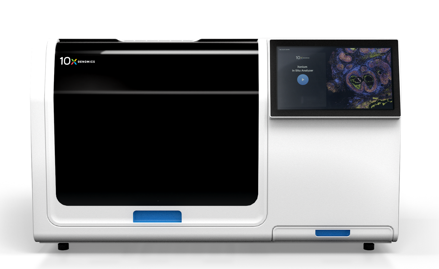
10x Xenium
High-Resolution In Situ Spatial Gene Expression Analysis
Customizable gene panels provide expression data at the tissue, cellular, and subcellular levels. The Xenium in situ spatial genomics platform enables simultaneous analysis of up to 5,000 genes using predefined panels, or up to 480 genes with custom-designed panels. This allows researchers to visualize gene expression within the context of tissue architecture, cell-cell interactions, and subcellular structures, revealing previously unseen biological mechanisms in both health and disease.
The extensive gene coverage supports the investigation of large transcriptomic networks and specific signaling pathways, including those related to immune function and cancer. With nanometer-scale imaging resolution, the Xenium platform integrates spatial gene expression data with traditional tissue stains such as H&E and immunofluorescence staining, enabling the identification of transcripts within specific cell types.
The Xenium Workflow
We can take your sample at any processing stage for our 10x Xenium lab services. However, it is important to remember that samples must be mounted on specialized Xenium slides for analysis. Our experts perform robust quality control (QC) on your sample, with H&E staining to ensure the morphological integrity of the tissue sample before analysis. We ensure accurate placement of high-quality tissue samples onto the Sample Area of Xenium slides.
High-Quality Sample Preparation
Precise Hybridization and Segmentation
Pre-defined or customized probe sets are added to the fixed sample and allowed to attach to their target RNA. After washing to remove excess probes, a ligase is added to anneal the complementary RNA to the circular probe, which is then enzymatically amplified.
Robust Segmentation, Analysis, and Further Processing
Cell segmentation is performed using different antibodies to label cell membranes, the cell interior, ribosomal RNA, and the nucleus. The Xenium Analyzer instrument generates an image-based signature of gene expression within each cell. The slide can be used for further H&E and immunofluorescence staining to generate more insights after analysis with the Xenium.
10x Visium
Unbiased Whole Transcriptome Single-Cell Analysis
Access whole-transcriptome, species-agnostic, FFPE 10x Genomics analysis of tissues to uncover spatially resolved gene expression profiles, splice variants, SNVs, and more. The Visium platform enables spatial analysis directly from tissue mounted on standard glass slides using probe hybridization with specialized Visium HD slides. It integrates seamlessly into conventional histology workflows, whether you supply pre-stained slides or choose our histology services delivering deep insights into cells and tissues of interest while supporting unbiased discovery.
The Visium is an ideal starting point for a wide range of research projects, offering comprehensive transcriptome coverage across complex tissue types in a single experiment. This broad analysis supports immediate insights and enables researchers to continuously revisit the data later for deeper or more targeted analysis to unlock new areas of inquiry.
The Visium Spatial Transcriptomics Workflow
High-Quality Sample Preparation:
We can work with your samples at any processing stage for our 10x Visium lab services, from fresh biopsy to archival tissue sections already mounted and fixed on glass slides. To help ensure you are analyzing the correct area of interest, we offer H&E and immunofluorescence staining before analysis. We also offer secure long-term sample storage with efficient retrieval for any future analyses you may require.
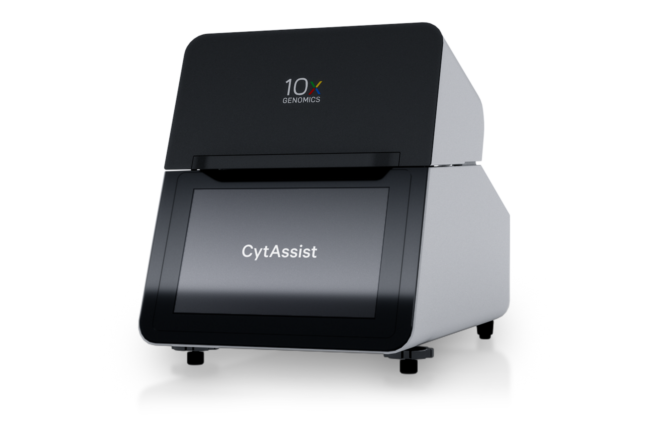
Advanced CytAssist Probe Capture and Barcoding
After staining, whole transcriptome probe panels are applied to samples. After hybridization and ligation of the probes to their targets, the CytAssist enables the transfer of the probes to a Visium HD slide, which contains millions of 2 x 2 µm barcoded squares.
Robust Library Preparation and Sequencing
A gene expression library is prepared for each square, and next-generation sequencing (NGS) is performed. The barcoded squares enable researchers to trace specific gene expression patterns to precise tissue areas. The expression data can be visualized as an overlay on H&E and immunofluorescence staining. Segmentation, guided by the 2 x 2 µm squares, enables resolution at the cellular and subcellular level.
Expert Data Analysis and Reporting
The whole transcriptome spatial analysis data retrieved from Visium analysis can be analyzed and formatted to match your requirements by our seasoned team of bioinformaticians. Alternatively, we can send you all of the data to analyze in-house in a format that best matches your internal workflows.

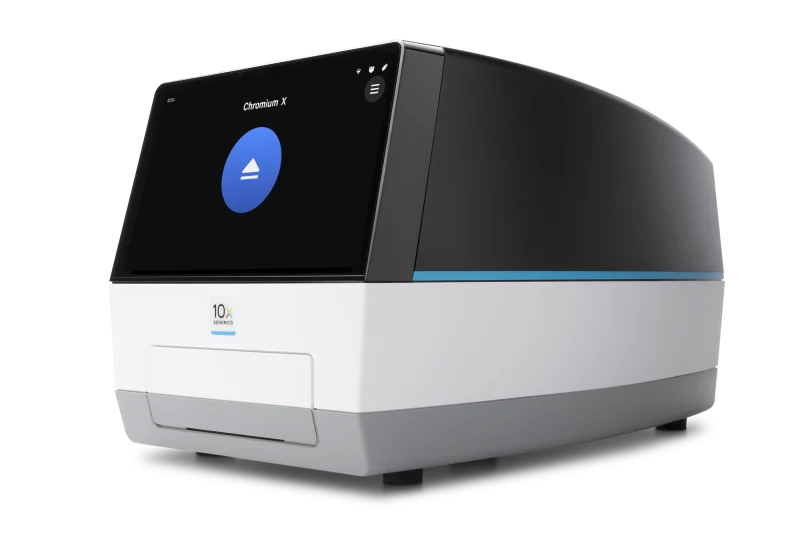
Single-Cell RNA and ATAC-Seq – 10x Chromium
High-Resolution In Situ Spatial Gene Expression Analysis
Gain precise single-cell transcriptomics insights using the best-in-class single-cell RNA seq (scRNA-seq) and single-cell ATAC-seq (scATAC-seq) platform, the Chromium from 10x Genomics. Chromium chemistry enables single-cell transcriptome profiling and the assessment of genome accessibility through ATAC-seq. The instrument leverages gel beads-in-emulsion technology, encapsulating single cells with sequencing reagents to enable rapid, high-throughput processing of thousands of single cells in a single run. The Chromium platform is compatible with spatial transcriptomics workflows, allowing researchers to map gene expression within a specific tissue for deeper biological insights.
The Chromium platform offers robust analytical capabilities to support advanced research in discovery and clinical contexts, such as detailed characterization of tumor microenvironments and immune cell infiltration. In clinical trials, single-cell analysis helps assess how different cell types respond to potential treatments.
The Chromium Workflow
Secure Sample Delivery and Preparation
We accept a wide range of sample types for our 10x Chromium lab services, including cell lines, primary tumor cells from patients, or cells derived from dissociated tissues. Whatever the source, we prepare the cell solution so it is ready for use in the Chromium instrument.
Single-Cell Analysis
Our team handles all wet lab work to ensure optimal single-cell data generation from the Chromium instrument. With deep expertise in using this technology, we’re fully equipped to address any challenges that may arise during the workflow. We utilize the automated Chromium Connect system to deliver consistent, high-quality performance and reliable sample processing at scale.
Analysis and Reporting
Our team of bioinformaticians are experts at analyzing and reporting advanced NGS experiments. We ensure you get the most out of your experiment and can provide any level of customization you require in the analysis. We also make sequencing and scATAC-seq data available in a format of your choice to match your internal workflows.
Integrated Workflows
These three instruments work excellently in tandem to achieve broad and deep insights into the tissue of interest. For instance, you could use the Xenium to assess a panel of disease-associated genes, such as the immuno-oncology panel, to see if your specific area of interest is impacted. If you get a positive hit, you could use the same tissue block and perform a more widespread analysis of the same tissue using the Visium to understand the broader transcriptome of different regions.
Conversely, you could begin with Visium or Chromium to obtain a broad overview of gene expression across the tissue. Then, using Xenium, you can focus on a specific, customized panel of genes to pinpoint their spatial expression, such as within the main tumor mass or at the interface with healthy tissue, to identify the cell types present in these regions and understand their roles.
Consistent End-to-End Processing
Our facility possesses all three 10x Genomics instruments. This means you can eliminate the need to send and receive your sample multiple times per analysis and can store it securely between runs, removing a major source of risk from your workflows and ensuring consistency in handling and processing.
Our histology services support the full range of processing and QC steps required for Visium and Xenium analyses, including embedding, sectioning, and H&E staining. You can rely on our experts to handle the most tedious and technically demanding parts of the workflow, ensuring you get the highest-quality results from your spatial genomics experiment.
Benefits of Using All Three 10x Genomics Platforms
Access to Cutting-Edge Platforms
We provide best-in-class instrumentation for our clients’ spatial omics studies, offering the Xenium, Visium, and Chromium from 10x Genomics. With these tools in your arsenal, you can answer pressing scientific questions and establish yourself as a leader at the cutting edge of any research field. You can transform your understanding of cellular function, discover novel cell types, and uncover targetable mechanisms with the power of 10x instrumentation. Furthermore, if you just want to run one assay, you do not need to invest capital in your own 10x instruments.
Expert Support and Customization
We provide the expertise to ensure you use 10x technologies to their full potential. Our experts understand the potential of spatial genomics and will ensure you maximize the information you receive from your sample. Furthermore, we can guide you through follow-up experimentation so you can fully capitalize on your results and drive your research forward.
Comprehensive Services Ensure Accuracy
We provide a full suite of services to support spatial genomics and ensure accuracy. We can arrange your sample shipping with custom kitting to ensure sample integrity. From there, we cover storage, processing, and QC within our extensive histology services. After analysis using a 10x instrument, we provide data analysis and reporting to ensure you receive high-quality, interpretable results that drive meaningful biological insights and support your next steps.
Scalable to Your Needs
We support projects of any scale, from early-stage studies with limited sample numbers to large cohort analyses, enabling targeted gene investigations and unbiased discovery research. Our services adapt to your needs, enabling the scaling up of analysis and the diversification of research into areas across cell biology and multiomics.
Leverage Spatial Genomics Technology Across Diverse Research Areas
Our advanced spatial genomics platforms allow you to perform cutting-edge experiments across diverse discovery and clinical research applications.
- Diagnostics
- Drug discovery
- Pharmacogenomics
- Clinical trials
- Research studies
- Biomarker discovery
- Population health studies
- Novel genome assembly
- Agrigenomics
- Disease modeling
- Development modeling
- Drug testing
In-Depth Spatial Insights From a Broad Spectrum of Sample Types
Our services offer insights into a wide range of sample types. We accept samples at nearly any processing stage, so you can rely on us to support your project, no matter where you are in your workflow. We support studies using a variety of species, including human, mouse, and rat. The types of samples we can receive include:
- Tissue biopsies
- FFPE tissues
- Samples fixed to microscope slides
- Blood
- Plasma
- Serum
- Saliva
- Hair
- Nail
- Stool
- Urine
- Semen
- Cell line samples/pellets
- Buccal swabs
Find Out More About Our End-to-End Single Cell and Spatial Omics Services
Want to discover how you can advance your research with our single cell and spatial omics services? Contact a member of our team to learn how you can best use this advanced technology to drive your research